Abdominal Blood Vessels Labeled / Circulatory Pathways Anatomy And Physiology Ii - New blood vessel growth is called angiogenesis.. These vessels transport blood cells, nutrients, and oxygen to the tissues of the body. Not only do blood vessels carry oxygen and nutrients, they also transport carbon dioxide and waste products away from our cells. The blood vessels are the components of the circulatory system that transport blood throughout the human body. Blood vessels form the living system of tubes that carry blood both to and from the heart. Oxygenated blood is then returned to the left atrium of the heart by four pulmonary veins.
Blood vessels are vital for the body and play a key role in diabetes helping to transport glucose and insulin. Oxygenated blood is then returned to the left atrium of the heart by four pulmonary veins. Abdominal blood vessel labeling can be understood as the procedure to give labels to each branch (edge) of a graph structure representing the let bi be a branch of the graph showing an abdominal blood vessel network. Label the blood vessels and structures using the hints provided. Blood vessels of the upper limb.
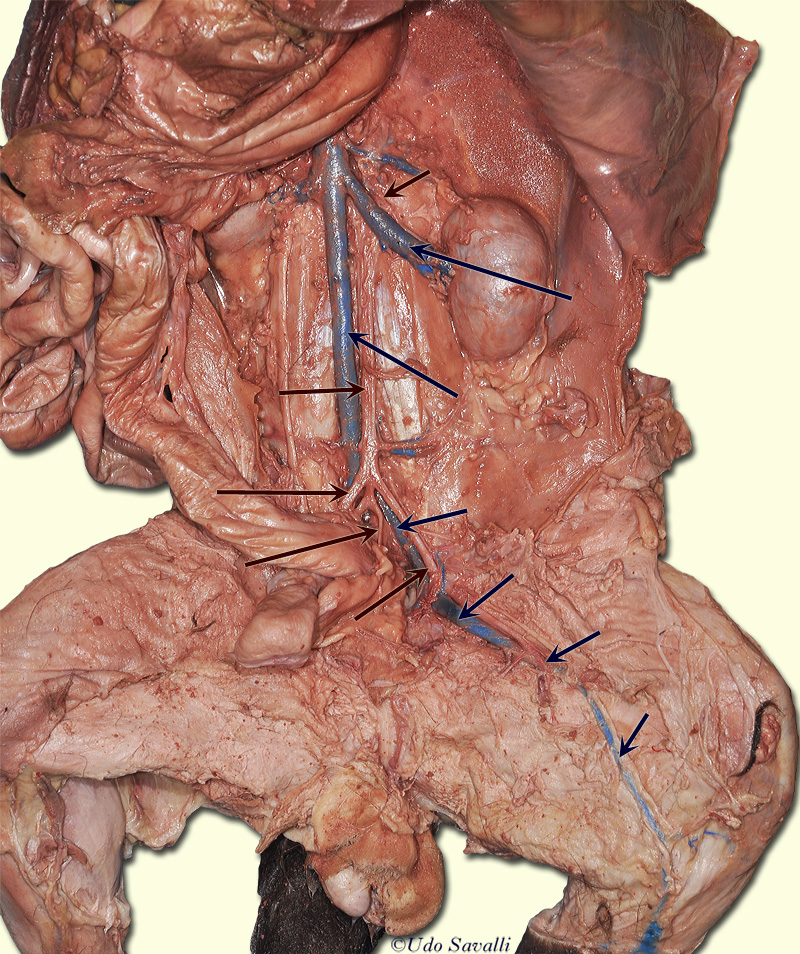
Bio202 Cat Vessels from www.savalli.us In abdominal surgeries, understanding blood vessel structure is critical since it is very complicated. Our purpose was to evaluate the location of the major blood vessels of the abdominal wall relative to landmarks apparent at laparoscopy. Label the steps in the homeostatic response to high blood pressure. Major blood vessels with pulmonary and systemic circuits. Nerves originating from lumbar region. Role of the use of omental flap in prognosis of cases with induced acute. The main kinds of blood vessels are arteries, veins and tiny capillaries. Dimitrios mytilinaios md, phd • last reviewed:
Our purpose was to evaluate the location of the major blood vessels of the abdominal wall relative to landmarks apparent at laparoscopy.
Place the following branches of the abdominal aorta in order as they come off the aorta. Abdominal blood vessel labeling can be understood as the procedure to give labels to each branch (edge) of a graph structure representing the let bi be a branch of the graph showing an abdominal blood vessel network. Role of the use of omental flap in prognosis of cases with induced acute. The blood circles the body around and around your whole life. They also take waste and carbon dioxide away from the tissues. This activity contains 12 questions. Blood vessels of the upper limb. .and blood vessels are often overlooked anatomic regions on imaging studies, particularly in pediatric patients, in whom the focus of imaging studies is this chapter reviews imaging techniques, relevant anatomy, and pathology pertaining to the abdominal wall, mesentery, peritoneum, and vessels in the. Blood vessels are referred to collectively as the vascular system and, together with the heart, make up the circulatory system or cardiovascular system. The blood vessels are the components of the circulatory system that transport blood throughout the human body. Posterior abdominal wall and blood vessels. Oxygenated blood is then returned to the left atrium of the heart by four pulmonary veins. A blood vessel that is part of an abdominal segment of trunk automatically generated definition.
Blood travels from the heart in arteries, which branch into smaller and smaller vessels, eventually becoming arterioles. Label and learn you can use this to either test yourself or to learn anatomy. Human anatomy for muscle, reproductive, and skeleton. The blood vessels of the body form a circle that begins and ends at the heart. There is a printable worksheet available for download here so you can take the quiz with pen and paper.
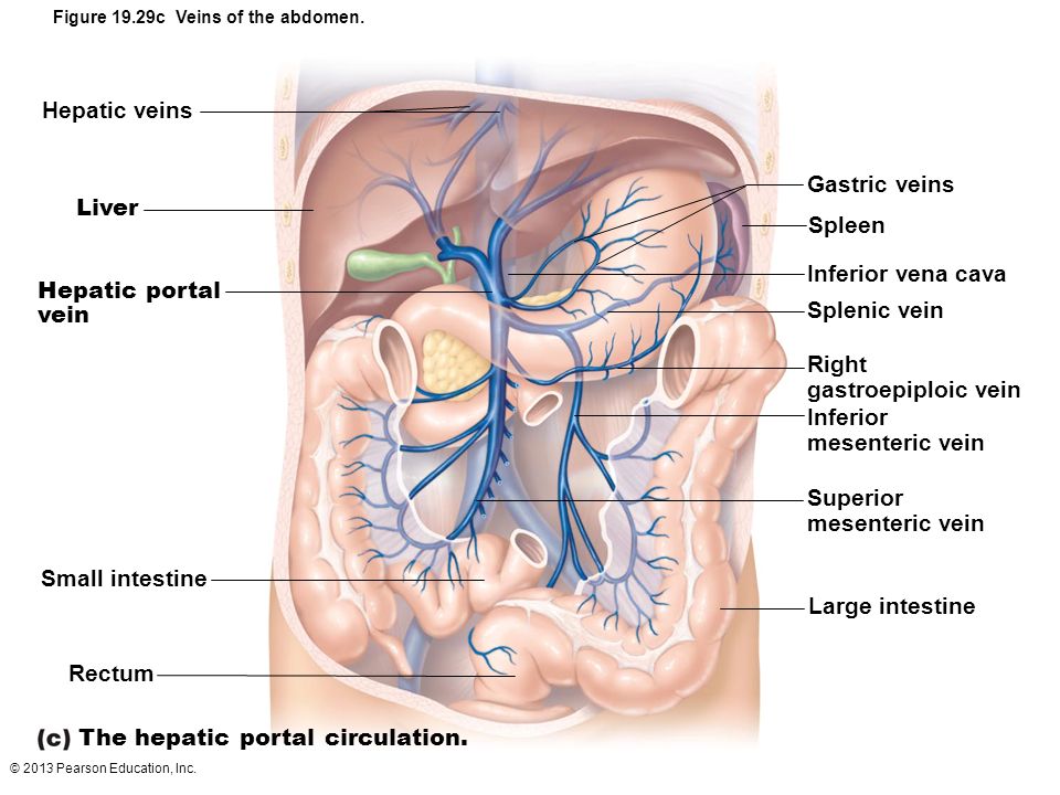
33 Label The Veins Of The Hepatic Portal System Labels Design Ideas 2020 from slideplayer.com New blood vessel growth is called angiogenesis. It includes all the arteries covered: The intestines have very rich blood supply. Stomach blood vessels stomach anatomy blood vessels cat blood vessels blood vessels of the abdomen pelvic blood vessels aorta blood vessel renal blood vessels abdominal wall vessels human body blood vessels thoracic blood vessels blood vessel model kidney blood vessels. Blood vessels of the upper limb. As a medical student, i found anatomy pretty challenging. The five types of blood vessels are (in order of circulation): Major blood vessels with pulmonary and systemic circuits.
Blood vessels form the living system of tubes that carry blood both to and from the heart.
Blood is oxygenated in capillaries that flow through the alveoli of the lungs. The thoracic aorta supplies blood to viscera of the. All cells in the body need oxygen and the vital nutrients found in blood. Dimitrios mytilinaios md, phd • last reviewed: An arterial, venous, or portal venous network can be represented by a tree. The blood vessels of the body form a circle that begins and ends at the heart. Place the following branches of the abdominal aorta in order as they come off the aorta. Blood vessels can be damaged by the effects of high blood glucose levels and this can in turn cause damage to organs, such as the heart and eyes, if significant blood vessel damage is sustained. Pictures and 3d models played a great role in helping me learn anatomy. Blood vessels of the upper limb. Many algorithms have been developed to accurately. .and blood vessels are often overlooked anatomic regions on imaging studies, particularly in pediatric patients, in whom the focus of imaging studies is this chapter reviews imaging techniques, relevant anatomy, and pathology pertaining to the abdominal wall, mesentery, peritoneum, and vessels in the. New blood vessel growth is called angiogenesis.
New blood vessel growth is called angiogenesis. Blood is made of cells and plasma. Nerves originating from lumbar region. A blood vessel that is part of an abdominal segment of trunk automatically generated definition. Arteries, arterioles, capillaries, venules, and veins.
Inferior Phrenic Arteries Wikiwand from upload.wikimedia.org Label the blood vessels and structures using the hints provided. Blood vessels can be damaged by the effects of high blood glucose levels and this can in turn cause damage to organs, such as the heart and eyes, if significant blood vessel damage is sustained. Role of the use of omental flap in prognosis of cases with induced acute. They are vital for carrying nutrients, oxygen and waste around the body. As a medical student, i found anatomy pretty challenging. The five types of blood vessels are (in order of circulation): Abdominal blood vessels labelled on gross anatomy specimen. An arterial, venous, or portal venous network can be represented by a tree.
Arteries, arterioles, capillaries, venules, and veins.
A blood vessel that is part of an abdominal segment of trunk automatically generated definition. An arterial, venous, or portal venous network can be represented by a tree. 1) starts at entry into abdominal cavity through aortic hiatus of diaphragm and ends by bifurcating at level l4 vertebrae into right and left common iliac arteries a) runs down midline of abdominal cavity; The input of the proposed method is the blood the anatomical labeling of blood vessel branches is performed by maximum a posteriori estimation. Blood vessels of the upper limb. Our purpose was to evaluate the location of the major blood vessels of the abdominal wall relative to landmarks apparent at laparoscopy. Major blood vessels with pulmonary and systemic circuits. Role of the use of omental flap in prognosis of cases with induced acute. The abdominal wall has quite a few blood vessels. Blood vessels can be damaged by the effects of high blood glucose levels and this can in turn cause damage to organs, such as the heart and eyes, if significant blood vessel damage is sustained. Human anatomy for muscle, reproductive, and skeleton. Abdominal blood vessels labelled on gross anatomy specimen. Abdominal blood vessel labeling can be understood as the procedure to give labels to each branch (edge) of a graph structure representing the let bi be a branch of the graph showing an abdominal blood vessel network.
We applied the proposed method to 50 cases blood vessels labeled. Molly smith dipcnm, mbant • reviewer:


0 Komentar